Brain
General program in the brain
Impact, the Dirk Hamer Syndrome (DHS)
A Hamer Focus (HH) appears in the brain.
CA phase or Conflict Active phase (stress phase)
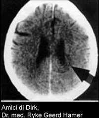
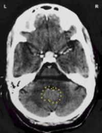
Concentric circles are visible on the CT scan, as if a pebble was thrown into the water.
It is an energetic event: something has passed by and a trace is visible, like footsteps in the snow.
In the center of the concentric rings, a meaningful biological special program is now initiated (biological association, see first biological law).
Resolution or conflictolysis (CL)
Production of cerebral edema in the HH.
PCL phase A or Postconflictolysis phase A (recovery phase A)
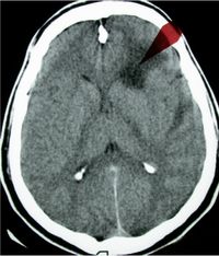
Brain edema develops at the location of the concentric rings. Dark spots can now be seen on the CT scan.
Epileptic or epileptoïd crisis (EC).
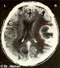
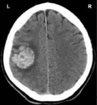
The edema in the HH is pressed out and it starts to fill with glia tissue (connective tissue in the brain). This happens from the outside in. The glia tissue is better perfused and may become visible on a CT scan as a light or white ring on the outside of the HH. This is often diagnosed as a brain tumor.
The crisis can also be diagnosed as a brain hemorrhage or cerebral infarction. Whether it is seen as a brain tumor, cerebral hemorrhage or cerebral infarction depends on what is seen on the scan, the symptoms and other physical symptoms or diagnoses.
PCL phase B or Postconflictolysis phase B (recovery phase B)
The HH fills up with glia tissue.
At the end of the program, a CT scan shows a lighter spot. This can be seen as scar tissue and is in indication that the program has come to an end. This, too, can be diagnosed as a brain tumor.
If the conflict is no longer triggered, this bright spot will integrate in the overall brain tissue and will become less clear.
All symptoms and diagnoses in the brain during and after the program are related to the activities of the Hamer Focus.
End program
After the program has finished, the Hamer Focus remains visible as a washout pattern.
Structure
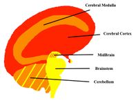
The brain can be divided into two parts, and each part can also be divided into two parts, see also the picture below:
- Old brain, consisting of the brainstem, midbrain and cerebellum
- New or large brain, consisting of the cerebral medulla and cerebral cortex
Function and programs
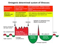
The function of the old brain and how it controls its associated organs and tissues is very different from that of the new brain. They are also derived from a completely different period in our evolutionary development. The illustration below shows the general characteristics of the programs in the old brain and in the new brain.
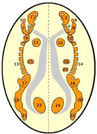
Relays (control centers) of organs and tissues in the brainstem
In the structure of the relays in the brain stem, the old ring-shaped animal can still be found, also see also endoderm.
1: Mucosa right side head and throat
2: Lung alveoli, O2 intake
3: Pharyngeal submucosa lower 1/3rd
4: Stomach
5: Mid ear right
6: Iris right eye
7: Lever
8: Pancreas
9: Duodenum
10: Smal intestines, jejunum
11: Kidney collecting tubes right kidney
12: Kidney collecting tubes left kidney
13: Small intestines, ileum
14: Appendix, colon ascending part
15: Colon, transverse part
16: Colon, descending part
17: Iris left eye
18: Mid ear left
19: Colon, sigmoid
20: Bladder trigone
21: Lung alveoli CO2 excretion
22: Mucosa left side head and throat
23: Left uterus, fallopian tube and prostate
24: Right uterus, fallopian tube and prostate
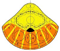
Relay of organs and tissues in the cerebellum (cerebellum)
Starting from the cerebellum, the brain and organs are crossed, also see old mesoderm. That is, the left side of the body is controlled by the right hemisphere of the brain and vice versa. Thus, the concentric rings of the worry-struggle-nest conflict manifesting itself in the right breast are on the left side of the brain (1) and that of an attack conflict, which manifests on the left side of the abdomen, is on the right side of the brain (6).
1. Breast glands right breast
2. Beast glands left breast
3. Corium skin left side body
4. Corium skin right side body
5. Peritoneum/omentum right side body
6. Peritoneum/omentum left side body
7. Right pericardium
8. Left pericardium
Relay of organs and tissues in the cerebral medulla
The relays in the cerebral medulla control the bones, tendons, muscles, joints, connective tissue, fat tissue, lymph nodes and vessels, and blood vessels at a particular location in the body. The exact content of the self devaluation conflict determines which body part is affected and which tissue in that body part, see new mesoderm.
1. Scull, right side
2. Scull, left side
3. Neck, right
4. Neck, left
5. Right hand and fingers
6. Left hand and fingers
7. Right arm and elbow
8. Left arm and elbow
9. Right shoulder
10. Left shoulder
11. Left myocardium
12. Right myocardium
13. Back, right side
14. Back, left side
15. Adrenal cortex right side
16. Adrenal cortex left side
17. Right hip
18. Left hip
19. Right Knee
20. Left knee
21. Right foot and toes
22. Left foot and toes
23. Right testicle/ovary-left kidney
parenchyma (2 cm down)
24. Left testicle/ovary-right kidney
parenchyma (2 cm down)
25. Speen (2 cm down)
Exceptions
- Because of the evolutionary rotation of the heart, the left heart muscle is controlled by the left hemisphere and the right heart muscle is controlled by the right hemisphere of the brain.
- Controle of kidney parenchyma is not crossed: parenchyma left kidney is controlled by the left hemisphere and that of the right kidney by the right hemisphere of the brain.
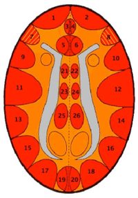
Relay of organs and tissues in the cerebral cortex
Also see ectoderm.
1. Thyroid ducts and pharyngeal ducts
right side
2. Thyroid ducts and pharyngeal ducts
left side
3. Tooth enamel right side mouth
4. Tooth enamel left side mouth
5. Alpha islet cells pancreas
6. Beta islet cells pancreas
7. Innervation laryngeal muscles
8. Innervation bronchial muscles
9. Laryngeal surface mucosa
10. Bronchial surface mucosa
11. Coronary veins, menstruation,
ovulation, cervix, vagina
12. Coronary arteries, penis/clitoris,
seminal vesicle + ducts
13. Rectum surface mucosa
14. Gallbladder, liver and pancreas ducts
15. Renal pelvis, bladder, urinary tracts
right side
16. Renal pelvis, bladder, urinary tracts
left side
17. Left halves retina body both eyes
18. Right halves retina body both eyes
19. Left halves vitreous body both eyes
20. Right halves vitreous body both eyes
21. Muscle innervation right leg and foot
22. Muscle innervation left leg and foot
23. Upper skin right leg and foot
24. Upper skin left leg and foot
25. Periosteum right leg and foot
26. Periosteum left leg and foot
Exception regarding the retina and vitreous body of the eyes
Left and right is not about the left or right eye but the left or right halves of the retina or vitreous body in both eyes.
These are also not crossed, because the left halves of both the vitreous body and the retina are controlled by the relay in the left hemisphere of the brain.
However, the left half of both eyes look to the right and the right half to the left. The one being looked at matters: mother/child or partner/3rd person, see biological laterality.
Evolution and parts of the cortex
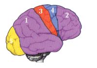
The cortex is divided is 5 parts:
- Post-sensory Cortex
- Pre-motor sensory Cortex
- Sensory Cortex
- Motor Cortex
- Visual Cortex
Before the ring shape broke, there were only 2 parts of the cortex: the post-sensory cortex and the pre-motor sensory cortex. They joined together and were one. After the ring broke, the motor cortex and the sensory cortex were inserted to controle the new tissues of the outer skin and the muscles.
More information about this topic during the online workshop "Bio-Logical Laws Completed"

